Brain MRI Viewer is a local workstation I built to explore my own scan data from a research study I participated in. It opens NIfTI volumes in an interactive multiplanar viewer, calculates deterministic file and signal checks, overlays a FastSurfer anatomy map, and keeps every research comparison tied to its method and limitations.
Public showcase: mri.shawnz.info. The hosted version retains the T1’s 256 × 256 × 208 matrix and the complete 64 × 64 × 33 × 240 resting-state series. It uses header-stripped, intensity-quantized derivatives and brain-masks the T1 rather than publishing the untouched source files.
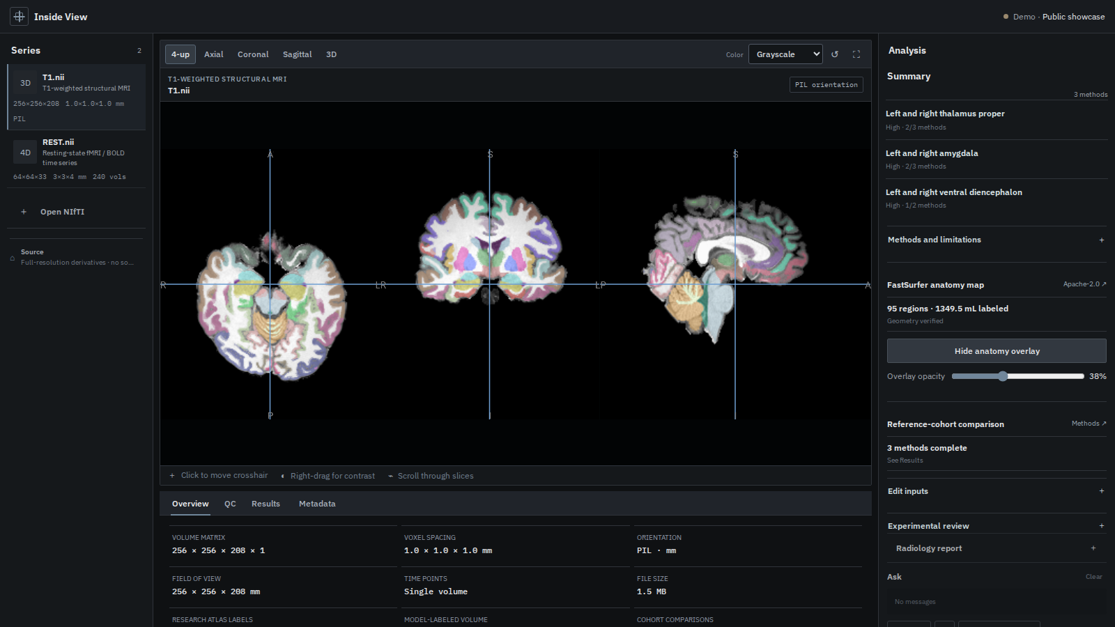
Implemented
- Synchronized axial, coronal, sagittal, multiplanar, and 3D NIfTI views.
- Frame navigation plus tSNR, DVARS, drift, and outlier summaries for 4D resting-state data.
- Local FastSurferVINN segmentation with a 95-region inspectable atlas overlay.
- Fixed-volume, PCN Braincharts, and CentileBrain comparison adapters with provenance and explicit compatibility labels.
- A consolidated Results view that reports repeated signals across methods without presenting them as a diagnosis.
- Optional local MedGemma review and report-grounded chat. Scan bytes remain on the workstation.
The boundary matters
This is research and educational software, not a medical device. A single T1 sequence cannot replace a complete MRI protocol or radiology interpretation. Missing scanner metadata stays missing rather than being filled with population defaults, and cross-pipeline comparisons are labeled exploratory.
The public deployment is deliberately narrower than the local application: full-resolution viewing and precomputed results are available, while segmentation, model inference, chat, and source-volume downloads are not.